Gallery

Team Member Name
Contact Bubble Networks
Elaina Lewis
This analogy represents contact networks as bubbles that leak viral particles. It describes how contact networks are not as small as they are perceived, and viruses can be spread unknowingly outside of the bubble.

Team Member Name
Candida albicans chlamydospore
Melina Woods
Unavailable

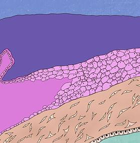
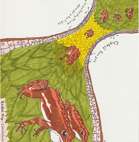
Team Member Name
Lemur Leap frog infected with chytrid fungus
John Lee
The Lemur Leap frog infected with the chytrid fungus Batrachochytrium dendrobatidis (Bd) has a ready defense in the dermal granular glands. The glands release a steady stream of antimicrobial peptides that can effectively kill the pathogen on the surface of the skin.

Team Member Name
Untitled
Shubhanjali Minhas
This cover art was created for a CRISPR/Cas9 system that the Nobile lab was working on. The piece likens a block tower to the genome of Candida auris, a fungus highly resistant to antifungal treatment. A Cas9 molecule is represented in the piece along with guide RNA. When this Cas9 molecule is used to take out certain genes, represented by individual blocks, in the genome, the block tower eventually falls, representing the heightened susceptibility of Candida auris to antifungal treatment. The light blue glow in the background represents the nucleus of Candida auris cells, while the orange globular shapes in the back represent the Candida auris cells.

Team Member Name
Introduction to the MERFISH Technology
Pinar Caglayan
My artwork consists of modular PNG drawings that aim to explain the MERFISH technology in a simple and clear way. Using these images, I put together a presentation that could be used by Dr. Christina Baer’s lab when introducing this technique to an audience that has no prior exposure to MERFISH.
Team Member Name
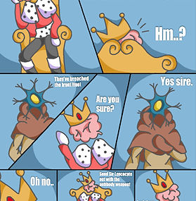
Untitled
Lauren Wong
This is the first comic panel of Sir Leucocyte's adventures against the deadly pandemic virus, COVID-19. In this scene, a neuron messenger walks into the throne room, "The head," and informs King Brain that the COVID-19 virus had broken through the first defense and has already laid waste to multiple healthy cells. King Brain informs the neuron messenger to send out our knight, Sir Leucocyte, with the antibody weapon.

Team Member Name
Untitled
Daphne Zhu
The gut microbiome is incredibly diverse and influences many aspects of health, many of which are yet unknown.

Team Member Name
Untitled
Erika Noda
Unavailable

Team Member Name
Activated macrophage
Anna Bright
Activated Macrophage

Team Member Name
Untitled
Emily Kopec
A battle in the colon between C. difficile and a neutrophil. C. difficile secretes toxins that release home into the gut; it is then able to repurpose that heme into a shield to protect itself. Neutrophilis (the green character) releases antimicrobial compounds at C. diff, but the shield renders them ineffective.

Team Member Name
Untitled
Eddie Qian
Unavailable

Team Member Name
Untitled
Zainah Siddiqi
"Rampant" is an oil painting focusing on the cell to cell communication and deterioration of the lungs when a person is infected by SARS-CoV-2. The background illustrates how people from all walks of life were impacted by the pandemic.

Team Member Name
Untitled
Michelle Kwon
Acinetobacter baumannii is a unique bacteria that tends to be widespread in hospital ICUs and surface tops. Rather than becoming weaker without the presence of water, it actually expresses the protein DtpA which allows the bacteria to tolerate environmental conditions without water. This piece depicts the bacteria in the center, glowing, as it expresses its unique protein while being surrounded by a body of water. The water that wraps around the bacteria is distant from the body representing the strength of the bacteria even without the direct presence of water. Furthermore, the blue water-like structure plays a dual role as it also represents the folded DtpA protein structure. Surrounding this entire figure are other bacterias that would usually need water and the presence of moisture in order to survive.

Team Member Name
Untitled
Liyan Shen
Unavailable

Team Member Name
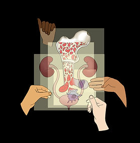
Hooked
Brigitte Jia
Renal T-cell on a fishing hook, above its parent organ, the kidney.

Team Member Name
Untitled
Justin Edaugal
A mixed media piece representing concepts of nutrition and bacteria. Portrait of a Filipino farmer holding a basket of fruit, representing nutrition, made using graphite pencil on paper. The portrait was then cut and layered with colored paper beneath to represent microbial dynamics. The Filipino farmer portrait is a nod to Filipino heritage of the David Lab Principal Investigator and the artist, myself. Graphite pencil, colored pencil on cut and layered, paper.

Team Member Name
Virus Abstract
Kadeer Wellington
This piece captures the stepwise assembly of a herpesvirus virion from the perspective of an assembly manual. The viral capsid is assembled in the nucleus of infected cells and is composed of distinct parts. This component will be combined with the viral genome. The second complex is prepared within the cytoplasm consisting of a lipid envelope containing viral proteins. The final steps involve combining the assembled capsid with the assembled viral lipid envelope to produce an infectious particle. Each part is digitally drawn and edited using MediBang Paint Pro.

Team Member Name
MFehi macrophages and MFelo macrophages
Liyan Shen
The mechanism of macrophages when there is
excess iron in adipose tissue in a lean setting.

Team Member Name
Brain poster
Aimee Li
My contributions involved a poster and two handouts. The microscope poster was designed for middle school students especially affected by COVID-19. Both the handouts are related to a brain activity for the same middle schoolers.

Team Member Name
Tree of Life
Melina Woods
This tree of life shows the relationship between different fungal species that form chlamydospores.

Team Member Name
Interconnectedness (black)
Navya Thakkar
This piece depicts the internal parts of the body that play a role in the inflammatory process of hypertension. Through abstract visualization of the body, it shows the link between DNA and the circulatory system, including arteries, veins and the heart. The immune cells (T-cells and dendrite cells) are shown to aggregate around the heart and the kidneys, causing inflammation in the organs, as well as play a large role in causing hypertension, shown by the arm cuff.

Team Member Name
Untitled
Elsa Runquist
This is a schema describing the process for how copy-back defective viral genomes (cbDVGs) are formed and their impact on the physiology of the cell, along with the relationship to respiratory syncytial virus (RSV) disease severity in infants. This illustration was initially made in BioRender, and then it was replicated using Photoshop drawing tools. (This is the Photoshop version).

Team Member Name
Ascano lab logos
Debbie Wang
These are the two logo designs that I created for the Ascano lab, each in three different color schemes. Both logos highlight the Acano lab's research focus -- the impact of viral infection on RNA-binding protein expression. Although based in biology, both logos are creatively designed with bold patterns and unique shoutouts. All DNA and RNA bases, for example, follow the conventional color scheme; however, both logos incorporate references to Nashville. The RNA strand on the left - designed to mirror a tangle of Christmas lights - include a G note in place of its m7G cap, as an homage to the music city. The RNA strand on the right includes hair pin structures shaped into the Nashville skyline. Through these logos, I hope to convey the interdisciplinary use of art in science.

Team Member Name
Labeled diagram of PIGR moving IGA through epithelial cells
Dayana Espinoza
Unavailable

Team Member Name
Untitled
Varvara Folimonova
These four drawings illustrate different types of placentas; epitheliochorial, synepitheliochorial, endotheliochorial, and hemochorial.
Team Member Name
Microglia devouring his plaque
Rina Shou
In both abstract art pieces, an amyloid beta plaque touches a microglia cell, which activates a signaling pathway that is involved in the removal of neurotoxic agents. Eventually, the microglia eats the plaque, which limits the progress of Alzheimer's Disease.

Team Member Name
Abscess Cartoon
Zhizhu Zhang
This cartoon depicts the immune cells and bacteria during abscess formation. Some neutrophils grab the magenta-colored S. aureus as a representation of phagocytosis. Others shoot arrows at the bacteria, which shows the process of exocytosis/release of ROS and proteases. Some dead neutrophils are also contained within the abscess, undergoing NETosis to trap the bacteria as well. The abscess is then encapsulated with fibrous and macrophages, which is represented by macrophages holding the barricade tapes at the perimeter. The cartoon uses the actual microscopic of the abscess formation process in the background. Zhizhu hopes to capture the dynamic interaction between different host immune components and invading microbes while making science fun and accessible.

Team Member Name
Untitled
Anjali Kumari
The image represents the HIV-HCV co-infection by portraying HIV (blue viruses) and HCV (red viruses) entering the cityscape. The stone figures and monuments signify places where the co-infections are prevalent and the countries' effort to prevent infection. Countries were selected based on literature searches that contained graphical representation of areas that are highly impacted.

Team Member Name
Untitled
Emily Krueger
My piece is an animation on the progress of a Group B Strep infection through the fetal membrane in pregnancy. This infection and resulting inflammation of the fetal membrane can have dangerous consequences during pregnancy, including premature labor and possible infection of the fetus.

Team Member Name
Untitled
Justin Edaugal
This digital piece illustrates the David Lab's research on Graft-versus-Host Disease (GVHD). A probiotic was given to a mouse model to study its affects on microbe behavior in the gut. This drawing is inspired by a cross-sectional, microscopic image of a normal colon on the left, and an image of an inflamed colon on the right as well as the atomic structures involved in the research.

Team Member Name
Untitled
Skylar Cuevas
One of the Byndloss Lab's research focuses is on intestinal inflammation induced by S. Typhimurium, a Salmonella enterica serotype. Inflammation allows the pathogen to outcompete other microbiota in the intestines by increasing the amount of the amino acid aspartate in the gut. This piece provides a cartoon visual of S. Typhimurium making the conversion of aspartate to fumarate in order to better thrive in the gut.

Team Member Name
Untitled
Betty Barnett
Collage of an intestinal brush border. A brush border is a collection of tiny protrusions on the surface of intestinal cells, which exist to optimize nutrient absorption.
Made using recycled paper materials.

Team Member Name
Global City
Elaina Lewis
This visualizes a concept for a global city with famous landmarks from around the world. It is a comment on how globalization results in an increase in the spread of viruses throughout the population.
Team Member Name
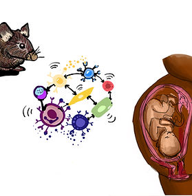
Untitled
Ereny Morcos
This summer, I worked in Dr. Nardhy Gomez-Lopez's lab to help generate a representation of the maternal-fetal crosstalk in humans and mice using single-cell technologies. My digital artwork showed the results received from a study the lab conducted where single-cell RNA-sequencing revealed unique cellular interactions in preterm labor that was driven by intra-amniotic infection.

Team Member Name
Untitled
Emily Layton
Unavailable

Team Member Name
Inactivated macrophage
Anna Bright
Inactivated Macrophage

Team Member Name
Untitled
Justin Edaugal
This digital piece illustrates a graphic comic-style, representation of the David Lab's research. What we eat can affect the concentration and type of microbes in our gut over time. Digital media.

Team Member Name
Ferroportin-1 Normal Vs. Dysfunctinal
Lucy Britto
Describes the normal function and mechanism of ferroportin, a multi-cellular iron exporter, compared to the influence of metabolic disease that may impact iron recycling via increased transporter degradation or mutations.

Team Member Name
Untitled
Martina D’Orso
The logo I created for Dr. Madhur’s lab is a sleek, contemporary logo that reflects all of their areas of studies combined with the words “Madhur Lab” to easily convey the name and meaning of the lab.

Team Member Name
Separations by Color
Stefan Marasligiller
Unavailable

Team Member Name
Phage in endosymbionts
Fei Yang
This work of the same medium zooms in on the Wolbachia bodies, and show the WO virus that contain genetic materials breaking out of the bacteria.

Team Member Name
Untitled
Daphne Zhu
Stool samples collected at three time points allows the David lab to learn about the diversity of microbes in participants' digestive tracts.

Team Member Name
Untitled
Audrey Kaul
This artwork was produced in collaboration with Lekha Nair based on her contributions to the work Mechanism of N6-Methyladenosine recognition by an RNA processing complex for driving IgH DNA recombination. The starting point of inspiration was drawn from the general idea of the coordination of multiple complex functions surrounding the decomposition and modification of RNA. The final artwork focuses on the process of decomposition through the placement of the m6A protein and its interaction with the nuclear m6A reader YTHDC1.

Team Member Name
Untitled
Laurence Gao
Unavailable

Team Member Name
Microglia devouring his plaque
Rina Shou
In both abstract art pieces, an amyloid beta plaque touches a microglia cell, which activates a signaling pathway that is involved in the removal of neurotoxic agents. Eventually, the microglia eats the plaque, which limits the progress of Alzheimer's Disease.

Team Member Name
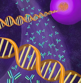
DNA, Antibodies, B Cell
Nadia Kafil
This piece depicts a B-cell with its DNA spilling out along with a wave of antibodies flowing through. The piece places a heavy emphasis on the DNA and antibodies of the B-cell which are arranged in a crisscrossing formation to represent the intersection of B-cell genome modification and its role in creating distinct antibodies.

Team Member Name
Untitled
Xinrui (Bella) Li
Unavailable

Team Member Name

Team Member Name
Obesity triggers
macrophage activity
Anna Bright
Abstract of adipose tissue invasion by immune system

Team Member Name
Untitled
Skylar Cuevas
Another research focus of the Byndloss Lab includes the connection between a high fat diet and E. coli choline catabolism. A high fat diet increases the bioavailability of nitrate and host-derived oxygen, leading to an expansion of E. coli. E. coli depends on nitrate for choline utilization, concluding that a high fat diet triggers choline catabolism by E. coli. This piece depicts a simplified version of the process beginning with diet and ending with catabolism in the gut.

Team Member Name
Untitled
Alexa Marcus
This is a small acrylic painting that depicts the A. baumannii bacteria, the central topic of my other pieces for the Skaar lab.
Team Member Name
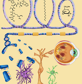
Untitled
Ellen Yu
Three conditions are commonly associated with diabetic retinopathy: high blood pressure, high fat, and high glucose. These conditions are represented by the chemical structures of angiotensin, palmitic acid, and glucose respectively. These factors impacted gene expression, so I inserted them inside loops of DNA, similar to the function of histones. This led to phenotypic changes in mueller glia, astrocytes, and microglia resulting in an overall phenotype that closely resembles diabetic retinopathy.

Team Member Name
Untitled
Diana Espinoza
This poster is meant to highlight the necessity of having compassion during stressful times such as a major health crisis. Part of what I believe contributes to having a better understanding of how a pandemic affects our peers and society is through obtaining efficient data visualization and modeling which the Colubri lab has been dedicated in making more accessible through multidisciplinary means.

Team Member Name
Untitled
Lauren Wong
Unavailable

Team Member Name
Salt Sensitivity Cover Art
Justin Yu
Cover art for a manuscript submitted to the Circulation Research journal, which showcases the article’s focus on salt-sensitive hypertension and the role of inflammasomes. A salt shaker is seen suspended and pouring its contents onto a kidney and blood vessels, representing the tissues that mediate salt-sensitive hypertension. The flames represent the inflammation, while the embers are shaped as inflammasomes and lymphocytes.

Team Member Name
IRP-IRE System
Lucy Britto
Overview of the relationship between iron homeostasis and the iron responsive elements/iron regulatory protein (IRE/IRP) system that coordinates the uptake, export, and storage of iron through post-transcriptional control mechanisms.

Team Member Name
CDTb Zipper 2
Eve Moll
This piece, titled 'CDTb Zipper 2' was likewise inspired by the zippering motion of the binary toxin of Clostridium difficile, and can also be read from left to right. The protein body has a unique shape, but retains its symmetry as the pore forms. We enter this imaginary, colorful, microscopic world to explore how such a protein moves and operates.

Team Member Name
Happy Blood Drop: Uniting urology, nephrology, and hematology
Kritika Bisht
This artwork was created to be a logo for the urology-nephrology-hematology teaching grant. It depicts the three branches working together to promote training, mentorship, and collaboration in the form of a happy blood drop. The hematology is represented by the blood drop, nephrology is represented by the lungs, and urology is shown attached to the lungs.

Team Member Name
Untitled
Ayo Sanusi
Nutrition interaction
between bacterial pathogens and vertebrae
hosts

Team Member Name
Iron Transfer Experimental Set Up
Lucy Britto
Visual schematic of a 17-day experimental protocol performed by a postdoc in the Hasty Lab involving primary cell isolation to study to the transfer of iron between differentiated adipocytes and polarized macrophages.

Team Member Name
Untitled
Ereny Morcos
Unavailable
Team Member Name
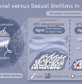
A Visual Comparision between Sexual and Conventional Biofilms
Anna Mehlhorn
In order to help audience members conceptualize sexual biofilms as they relate to conventional biofilms, I created a figure displaying both in the fungal species, Candida albicans. I intentionally used a soft color palette and cartoon-like illustrations to make an intimidating concept more approachable to the viewer.

Team Member Name
DVG Schema
Elaine Lewis
This is a digital schema of the different pathways defective viral genomes (DVGs) can undergo as they are integrated into a cell. DVGs can cause viral replication interference with the full viral genome. They could also interact with PKR and cause stress granules and antiviral immunity. Finally, they could undergo the RLR/MAVS signaling pathway and cause cell survival/persistence, antiviral immunity, or inflammation.
Team Member Name
Untitled
Erin Lee
The task was to create a urology-nephrology-hematology teaching grant logo. It was requested that the interaction between the three branches were highlighted, along with an emphasis on collaboration, mentorship, training, and diversity.
The goal was achieved by showing the different branches on separate slides and having different hands come together to create the final illustration.

Team Member Name
Untitled
Shubhanjali Minhas
This is another version of the CRISPR/Cas9 cover art. Here, the block tower is represented in a "zoomed in" environment, where a monochromatic blue color scheme is used to represent this event occurring in the nucleus. The background contains many strands of dna, further illustrating that this is occurring in the nucleus of a Candida auris cell.

Team Member Name
CcmA Distribution Patterns on Heliobacter pylori
Mariana Smith
This dynamic sculpture illustrates the 3D distribution of CcmA proteins on the cell wall surfaces of four H. pylori mutants (with each mutant type shown in a different color under natural light). When black light is introduced, the CcmA protein distribution on each cell becomes visible and begins to glow. Each cell shape is taken from a real cell in the lab, along with its corresponding protein distribution.

Team Member Name
Untitled
Elsa Runquist
This is a diagram describing the different techniques used by the Lopez Lab when studying copy-back defective viral genomes (cbDVGs). These techniques include VODKA, RNA FISH, and PCR. All three techniques are mapped out on a single page, allowing for greater efficiency when delivering this information to other students and researchers. This illustration was initially made in BioRender, and then it was replicated using Photoshop drawing tools. (This is the Photoshop version).

Team Member Name
Hypertension
Brigitte Jia
Cardiac and renal systems implicated in hypertension, depicted together.

Team Member Name
Untitled
Helen Qian
A depiction of drug molecules binding onto the CFTR protein to remedy loss of function mutations in cystic fibrosis.
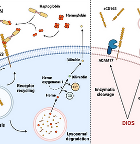
Team Member Name
CD163 Cycle Normal Vs. Dysfunctional
Lucy Britto
Comparison of the normal function of iron handling receptor CD136 and how metabolic dysfunction may impact CD163 shedding mechanisms such as enzymatic cleavage or extracellular vesicles.

Team Member Name
Untitled
Daphne Zhu
The Pediatric Obesity Microbiome & Metabolism Study (POMMS) utilized DNA extracted from stool samples to gain insights on the microbiomes of children with obesity. Using these DNA sequences, the David lab can also obtain a diet profile of foods that participants eat, which may be a more effective way to learn about individuals' diet histories than self-report, which can be time-intensive and is subject to recall bias.
This participant report returns the study's results as well as educational material on the microbiome to the individual that provided their stool samples.
Team Member Name
Cells, Antibodies, and DNA
Nadia Kafil
This piece depicts B-cells, antibodies, and strands of free-flowing DNA. These objects are intertwined with one another to represent the connection of these elements in the process of B-cell genome modification and the formation of unique antibodies.

Team Member Name
Untitled
Jessica Cascio
This summer I worked with the lacy Lab at VUMC to create cover art for two papers they are publishing. Through their research, the lab found uncanny similarities between the membrane proteins of two bacteria (one that infects the stomach and one that infects the lungs). The cover art I created serves to start a conversation about the similarities between these two complex structures.

Team Member Name
Untitled
Betty Barnett
Kaleidoscopic depiction of intestinal cells.
Derived from an image taken in the Tyska lab via freeze-fracture electron microscopy.

Team Member Name
Untitled
Diana Espinoza
This abstract work is meant to depict the rapidly evolving nature of mRNA viruses. The work contains motifs related to the replication of viruses in the body which are then used to compose a scene that mimics that of a city in chaos.

Team Member Name
ENaC-Mediated Salt Response
Justin Yu
Displays the mechanism of the ENaC-Mediated Salt Response, which plays a role in salt-sensitive hypertension.

Team Member Name
Untitled
Alexa Marcus
Mutations in the genome of A. baumannii, where ISAba11 is inserted upstream of ispB and reduces ispB;s expression, increases A. baumannii's cell size and antimicrobial resistance, making it more harmful. This is shown in that the bacteria are a deeper red as they become more virulent. As this transitions across the graphic, gene insertion and asymmetrical membrane restoration are depicted.

Team Member Name
Untitled
Daphne Zhu
We explored different ways to visualize the diet information obtained from stool samples.

Team Member Name
Untitled
Rachel Eom
An abstract rendering of the intersection between the renal (kidney), cardiovascular (heart), and immune (cytokines shown as white spheres) systems and how changes in the molecular machinery by DNA missense mutation, as well as changes in protein structure greatly influence hypertension conditions.
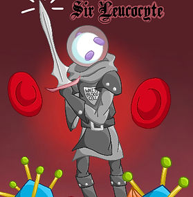
Team Member Name
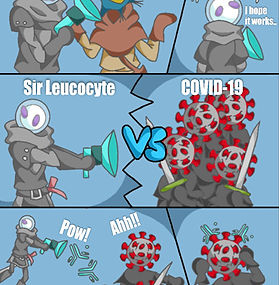
Untitled
Lauren Wong
This is the cover art to the short comic, "Sir Leucocyte," a story about a brave white blood cell knight who fights against various viruses to protect his kingdom, the Body.

Team Member Name
Untitled
Justin Yu
This piece of cover art depicts an anthropomorphized virion wearing a suit with a head shaped like an icosahedron, representing the Kaposi's Sarcoma-associated Virus (KSHV), a major focus of the Karijolich Lab. Jutting out from the back of the virion are a number of different mechanical elements, which are used to signify the synthetic nature of viruses, as they rely on a host's cellular machinery in order to reproduce. One gear is being removed by a disembodied hand. This represents the Karijolich Lab's research into the many mechanisms of KSHV infection, including the relationship between ORF36 and ISGylation.

Team Member Name
CcmA Distribution Patterns on Heliobacter pylori
Mariana Smith
This dynamic sculpture illustrates the 3D distribution of CcmA proteins on the cell wall surfaces of four H. pylori mutants (with each mutant type shown in a different color under natural light). When black light is introduced, the CcmA protein distribution on each cell becomes visible and begins to glow. Each cell shape is taken from a real cell in the lab, along with its corresponding protein distribution.
Team Member Name
The Fetal Membrane: A Focus on the Fetal-Maternal Interface
Alyssa Glauser
A diagram identifying the layers of the fetal membrane and how they come into contact with the maternal blood and decidua.
Team Member Name
Biosphere
Sophie Stark
Unavailable

Team Member Name
Interconnectedness (grey)
Navya Thakkar
This piece depicts the internal parts of the body that play a role in the inflammatory process of hypertension. Through abstract visualization of the body, it shows the link between DNA and the circulatory system, including arteries, veins and the heart. The immune cells (T-cells and dendrite cells) are shown to aggregate around the heart and the kidneys, causing inflammation in the organs, as well as play a large role in causing hypertension, shown by the arm cuff.
Team Member Name
Untitled
Natalie Elliott
Unavailable
Team Member Name
Untitled
Lauren Wong
This is the first comic panel of Sir Leucocyte's adventures against the deadly pandemic virus, COVID-19. In this scene, a neuron messenger walks into the throne room, "The head," and informs King Brain that the COVID-19 virus had broken through the first defense and has already laid waste to multiple healthy cells. King Brain informs the neuron messenger to send out our knight, Sir Leucocyte, with the antibody weapon.
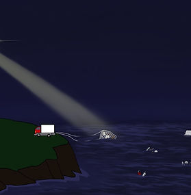
![This cartoon depicts a spaceship delivering the LTB4 lipids, using the actual microscopic image taken by the Serezani lab as background. The inspiration of spaceship was taken from an interesting mistake of the computer vision technology. When Dr. Serezani sent over some microscopic pictures for reference, Gmail described the pictures as "[A picture containing star, outdoor object, night sky, light Description automatically generated with low confidence]". Produced by macrophages and neutrophils, LTB4 is an important signaling mediator to enhance antimicrobial effector functions. However, excessive LTB4 can lead to aberrant inflammation that is damaging to host defense. Thus, some LTB4 molecules are portrayed to be devil-like.](https://static.wixstatic.com/media/33432f_c0e49ce5d29f4589b267bb7ae09893ae~mv2.jpg/v1/fill/w_279,h_285,al_c,q_80,usm_0.66_1.00_0.01,enc_avif,quality_auto/Image-empty-state.jpg)
Team Member Name
LTB4 Cartoon
Zhizhu Zhang
This cartoon depicts a spaceship delivering the LTB4 lipids, using the actual microscopic image taken by the Serezani lab as background. The inspiration of spaceship was taken from an interesting mistake of the computer vision technology. When Dr. Serezani sent over some microscopic pictures for reference, Gmail described the pictures as "[A picture containing star, outdoor object, night sky, light Description automatically generated with low confidence]". Produced by macrophages and neutrophils, LTB4 is an important signaling mediator to enhance antimicrobial effector functions. However, excessive LTB4 can lead to aberrant inflammation that is damaging to host defense. Thus, some LTB4 molecules are portrayed to be devil-like.

Team Member Name
Untitled
Varvara Folimonova
A topologically associating domain (TAD) is a region within the genome where genes are more likely to interact with one another. Dr. Basu's lab studies TADs along with other genetic elements within B cells to better understand their mechanism of generating antibodies.

Team Member Name
Untitled
Emily Layton
Unavailable
Team Member Name
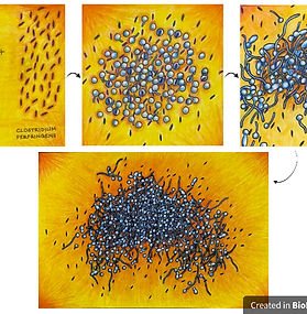
Untitled
Gayathree Gopi
My first piece depicts the formation of a special type of polymicrobial biofilm called a “mini-biofilm” between C. albicans and the anaerobic bacteria C. perfringens. The second piece illustrates the life cycle and process of mature mini-biofilm formation.

Team Member Name
Untitled
Gayathree Gopi
My first piece depicts the formation of a special type of polymicrobial biofilm called a “mini-biofilm” between C. albicans and the anaerobic bacteria C. perfringens. The second piece illustrates the life cycle and process of mature mini-biofilm formation.

Team Member Name
Untitled
Elsa Runquist
This is a schema describing the process for how copy-back defective viral genomes (cbDVGs) are formed and their impact on the physiology of the cell, along with the relationship to respiratory syncytial virus (RSV) disease severity in infants. This illustration was initially made in BioRender, and then it was replicated using Photoshop drawing tools. (This is the Photoshop version).

Team Member Name
Proteome study in pregnant women with COVID-19
Grace (River) Terrell
This piece shows the distinct effects in PC3 levels based on COVID severity and that infected pregnant women usually had a less severe case of COVID. The final element shows the larger cytokine storm and upregulation of angiogenic factors in non-pregnant individuals.

Team Member Name
Bar 3
Rebecca Dubin
This image depicts a concept that the Denison lab has been working on. One aspect that makes COVID-19 difficult to stop is that it has a proofreading mechanism protecting the replication of its RNA known as ExoN. It has been discovered that although 5FU, a common drug used to cause mutations, is blocked by ExoN, a drug known as Remdesivir is able to sneak around the ExoN bouncer.

Team Member Name
Genesis
Ardria McDonald
It represents how B cells develop, divide, and branch out to other organs from bone marrow, in which it all resembles a growing “tree”.

Team Member Name
Untitled
Varvara Folimonova
These four drawings illustrate different types of placentas; epitheliochorial, synepitheliochorial, endotheliochorial, and hemochorial.

Team Member Name
Karijolich lab logo
Justin Yu
This is a logo created for the Karijolich Lab. It includes a dark bar running through its center that is supposed to represent the black resin tops of lab benches. As seen in the logo, lab equipment commonly used in biomedical laboratories sits on top of this black bar. In the lower half, the "o" in Karijolich has been replaced with an icosahedron containing dsDNA, a representation of the Kaposi's Sarcoma-associated Virus, an important focus for the Karijolich Lab. Arrayed around the letters are a number of viral RNA structures that the lab studies as well.
Team Member Name
A Look Into Alzheimer's
Nishita Maknojia
This image showcases a healthy, normal brain besides an Alzheimer’s brain. The magnifying glass zooms into the diseased brain to highlight how microglia plays a role in AD pathogenesis by releasing inflammatory mediators which can contribute to amyloid beta plaque aggregation.

Team Member Name
Untitled
Skylar Cuevas
The Byndloss Lab's research centers on inflammation-induced gut dysbiosis and its role in non-communicable diseases such as IBD, cancer, and cardiovascular disease. As depicted in the piece, inflammation mediates an imbalance in the microbial ecosystem. A focus in the Byndloss lab is studying how inflammation changes result in gut dysbiosis.